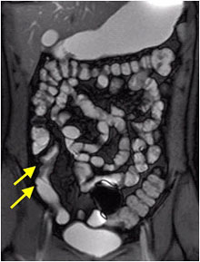
There is one precaution: no colonoscopy with electrocoagulation should be performed directly after the MRI because of methane resulting from Mannitol breakdown. Bethesda, MD 20894, Web Policies Colonoscopy plays a fundamental role in the diagnosis and monitoring of patients with CD. Crohn's disease is characterized by inflammatory lesions in the gastrointestinal tract, most commonly in the terminal ileum and colon. Ikhtaire S, Shajib MS, Reinisch W, Khan WI. MRE uses dynamic, high spatial resolution and soft tissue characterization of the bowel to provide vital anatomic and physiologic information without exposing patients to unnecessary ionizing radiation [5,6,7, 13]. Like standard colonoscopy, a thorough cleansing of the bowel is required beforehand. If a patient has a contraindication to receiving procedural anesthetic sedation, then a patient cannot undergo colonoscopy. If you have claustrophobia, or fear of confined spaces, or anxiety, your healthcare provider may give you a prescription for a sedative prior to your MR enterography. 189 patients with colonic CD were initially included in the study however 33 patients were excluded because of incomplete data. WebLike CT enterography, MR enterography uses a contrast agent to enhance images of the intestines. This helps your healthcare provider make a diagnosis, as well as monitor your treatment. Ugeskr Laeger. The median time interval to complete all exams was 8days. Skip lesions refers to the interspersed inflammation "skipping" parts of the bowel, which are left unaffected (green arrows). 2020;40(4):1020-1038. doi:10.1148/rg.2020200011, Saleh L, Juneman E, Reza Movahed M. The use of gadolinium in patients with contrast allergy or renal failure requiring coronary angiography, coronary intervention, or vascular procedure. Fecal calprotectin (FCP), magnetic resonance enterography (MRE), and colonoscopy are complementary biometric tests that are used to assess patients with Once all images are done, the exam table will slide out from the MRI tube. Sinha R, Rawat S. MRI enterography with divided dose oral preparation: Effect on bowel distension and diagnostic quality. Mayo Clinic Minute: What you need to know about polyps in your colon, Colon cancer screening Weighing the options, Advertising and sponsorship opportunities, The possible need for follow-up testing to investigate a false-positive finding or to remove tissue. The CIs for Pearsons were 0.610.77 while for Spearmans were 0.370.61 (Table4). Your healthcare provider will inform you where your exam will take place. 2017;15(1):5662. MRE compares favorably to colonoscopy for evaluation of known or suspected Crohn's disease noninvasively and without the exposure to ionizing radiation associated with CT enterography (CTE). MR enterography correlates highly with colonoscopy and histology for both distal ileal and colonic Crohn's disease in 310 patients The site is secure. Calafat M, Cabr E, Maosa M, Lobatn T, Marn L, Domnech E. High within-day variability of fecal calprotectin levels in patients with active ulcerative colitis: what is the best timing for stool sampling? Magnetic resonance enterography guiding treatment in children with Crohn's jejunoileitis. Measurements are best performed on the sequence with good luminal distension. FOIA  By Cathy Wong MR enterography: oral administration of contrast. Ordas I, Rimola J, Rodrguez S, Paredes JM, Martnez-Prez MJ, Blanc E, et al. Also, the colonoscope is not used if a person is too emotional to perceive the upcoming procedure and this affects his mental health. The CIs for Pearsons were 0.460.67 while for Spearmans were 0.620.78 (Table3). Cite this article. is a board-certified abdominal radiologist, currently working as an Associate Professor of Clinical Radiology in the Department of Radiology at the University of Texas Health Science Center at San Antonio where he is the Director of Abdominal Imaging. Cathy Wong is a nutritionist and wellness expert. The https:// ensures that you are connecting to the The P-values of the estimated Pearsons (rho=0.55) and Spearmans (rho=0.71) correlation coefficients were highly significant (P<.0001, respectively). By clicking Accept All Cookies, you agree to the storing of cookies on your device to enhance site navigation, analyze site usage, and assist in our marketing efforts. Magnetic resonance enterography (MRE) is a non-invasive medical imaging procedure that uses a magnetic field rather than ionizing radiation. Hartmann D, Bassler B, Schilling D, Adamek HE, Jakobs R, Pfeifer B, Eickhoff A, Zindel C, Riemann JF, Layer G. Radiology. This is sufficient for most therapeutic decisions. Bowel inflammation, fistulas and abscesses show restricted diffusion -high on DWI, low on ADC. We use a Mannitol in water solution (2%), which provides good contrast between lumen and bowel wall on both T1 and T2 sequences and is well accepted by patients. This content does not have an Arabic version. Crohns disease endoscopic index of severity, Health information portability and accountability act. Schreyer AG, Hoffstetter P, Daneschnejad M, Jung EM, Pawlik M, Friedrich C, Fellner C, Strauch U, Klebl F, Herfarth H, Zorger N. Acad Radiol. Virtual colonoscopy (VC), also known as computed tomography colonography, is an effective method for detecting polyps. CD activity on MRE was measured with the Magnetic Resonance Index of Activity (MaRIA). Tontini GE, Rath T, Neumann H. Advanced gastrointestinal endoscopic imaging for inflammatory bowel diseases. The colonoscope is also equipped with a device that allows you to immediately make a biopsy (take a sample) of tumors found in the intestine. At our institution, a pre-procedural oral preparation is used for bowel cleansing, which is essential for adequate mucosal examination. 8600 Rockville Pike WebThis procedure uses a magnetic field to create detailed images of your organs, instead of an X-ray or CT scan. Both T2-weighted images (HASTE and TrueFISP) and contrast-enhanced images show linear and transmural ulceration (12). Talk to your healthcare provider about how to proceed in the event of abnormal results. The results of the multivariate linear regression indicated that MaRIA scores (effect =1.54, P-value <0.0001) and CDEIS (effect=2.23, p-value <0.0001) had a significant and positive association with FCP levels (Table5, Fig. The future research should be directed towards streamlining the schedule and clinical decision of when to perform these exams. Achiam MP, Chabanova E, Lgager VB, Thomsen HS, Nielsen OH. Mayo Clinic does not endorse any of the third party products and services advertised. Epub 2013 May 3. Hospitalization and surgery occurred more in untreated patients than in subjects undergoing biological therapy (12.1% vs 0.0%, P = . 2019;37(7):5117. A fistulous track can present with a layered 'tram track' configuration or as a linear enhancing structure. In addition to regular preparation for an MRI exam, prior to MR enterography the patient is given two bottles of a special liquid to drink (one bottle 20 minutes before the exam and one bottle 10 minutes before the exam). information highlighted below and resubmit the form. A correlation between all three tests: FCP, MRE, and colonoscopy, has to our knowledge, never been shown in the same cohort of patients with colonic CD. Cerrillo E, Beltran B, Pous S, Echarri A, Gallego JC, Iborra M, et al. Elective surgery outcomes in inflammatory bowel disease: interpretation at magnetic resonance enterography. and transmitted securely. Before your procedure, you may want to ask your healthcare provider: In general, its also essential to understand why you are undergoing MR enterography. WebCharacteristic Radiologic Findings. CT enterography is a relatively new technique to evaluate Crohn disease. 2009;361(21):206678. MR enterography, also called Magnetic V.S.K. UCSF Department of Radiology and Biomedical Imaging. Certain patients may have contraindications to undergo MR imaging, such as medical devices, hardware, claustrophobia, or allergy to gadolinium contrast. The coronal T1 post-contrast image (left) and the T2 image (right) show skip lesions in the terminal ileum. Sensitivity was 92.04% (95% CI, 83.5897.21) and specificity was 85% (95% CI, 68.9193.48). VK performed critical revision of the manuscript, performed colonoscopic interpretations, and acquired data, which was analyzed and interpreted.VSK performed critical revision of the manuscript, performed radiological interpretations, and acquired data, which was analyzed and interpreted. This site needs JavaScript to work properly. It can be seen going from one bowel loop to another bowel loop, to another hollow organ or to the skin. At our institution, clinical disease activity was calculated for each patient at the time of MRE with the Crohns Disease Activity Index (CDAI) score.
  The image is a post-contrast T1 image with a mucosal enhancement pattern in the terminal ileum (arrow). Tell your healthcare team right away if you develop symptoms. https://doi.org/10.1186/s12876-019-1125-7, DOI: https://doi.org/10.1186/s12876-019-1125-7. During your check-in process, you may be asked to fill out a safety form. The test doesn't require bowel preparation, sedation or insertion of a scope. All rights reserved. In rare cases, other methods of research are allowed. The bowel is a common site for pathologic processes, including malignancies and inflammatory disease. FOIA This middle layer can consist of fat, edema or fibrotic tissue. Be sure to tell your healthcare team if you have any metal objects in your body. To get the best images, you must remain still and follow breath-holding instructions during the procedure. The affected lesions show increased enhancement with a layered pattern (yellow arrows), while another part is unaffected or skipped (green arrows). HHS Vulnerability Disclosure, Help Other indications include celiac disease, postoperative adhesions, radiation enteritis, scleroderma, small bowel malignancies, and polyposis syndromes. They should be aware of recent surgeries, as well as any medical devices, and implants. One hundred fifty-six patients with colonic CD were prospectively examined between March 2017 and December 2018. National Institute of Biomedical Imaging and Bioengineering. A prestenotic dilatation is seen before both segments. Colonoscopy For this procedure, a doctor inserts a thin, flexible tube with a video camera and light through the anus and into the rectum and colon. p.p1 {margin: 0.0px 0.0px 0.0px 0.0px; font: 8.5px Helvetica}
  Inclusion criteria were informed consent, 18years of age or older, known diagnosis of colonic CD, MRE performance, measurement of FCP levels within a maximum of two weeks prior to MRE, colonoscopy within a maximum of two weeks before or two weeks after the MRE, and no pharmacological therapy modification. MR enterography. Table2 presents the statistical analysis between FCP and CDEIS. Clipboard, Search History, and several other advanced features are temporarily unavailable. The P-values of the estimated Pearsons (rho=0.71, P<.0001) and Spearmans (rho=0.49, P<.0001) were highly significant. Anyone you share the following link with will be able to read this content: Sorry, a shareable link is not currently available for this article. showed that colonoscopy and MRE accurately assess healing of mucosal inflammation in the colon however FCP levels were not correlated [15]. Jensen MD, Nathan T, Rafaelsen SR, Kjeldsen J. Clin Gastroenterol Hepatol. In the grading system, only severe stenosis is included as a complication, which is defined as a stenosis with prestenotic dilatation and a moderate-to-marked increase in mural T2 signal. The .gov means its official. Your healthcare provider will then share these results with you. The results of the MRE were compared to the colonoscopy and pathology reports to determine the presence or absence of disease in evaluable bowel segments. 2012;3 (3): 251-63. Colonoscopy, in turn, if not painful, then a rather unpleasant diagnostic measure. MRE and US are well tolerated.
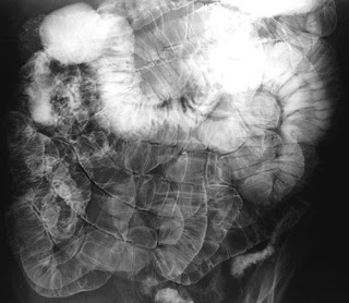
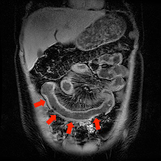
  At our institution, MRE is performed using 3.0 Tesla magnet systems (Siemens Healthcare, Berlin, Germany). This study was performed to compare findings of magnetic resonance enterography (MRE) and ileocolonoscopy. You may continue with your usual eating and drinking routine after the procedure. Abscesses are often seen in patients with severe active Crohn's disease. Check out these best-sellers and special offers on books and newsletters from Mayo Clinic Press. While prior studies have evaluated the association between combinations of these tests, no study has established a correlation between all three: FCP, MRE, and colonoscopy. Careers. Accessibility The procedure is carried out by the method of circular scanning, during which the person is located on a horizontal platform so that the area of investigation is inside the tomograph. Receiver operating characteristic (ROC) curve of fecal calprotectin (FCP) values to predict active disease on colonoscopy with CDEIS. Abraham C, Cho JH. When the colon is involved in Crohn's disease a decrease of haustral folds can be seen. 2015;21:15729. Sensitivity was 92.04% (95% CI, 83.5897.21) and specificity was 83% (95% CI, 67.9192.31). MRI is particularly useful for evaluating a fistula around the anal area (pelvic MRI) or the small intestine (MR enterography). The first available colonoscopy was considered as baseline, and patients were monitored over time to evaluate both the recurrence of disease postoperatively and the onset of negative disease outcomes. Unlike a CT scan or X-ray, MR enterography: One of the limitations of MR enterography is that movement may impact the image quality. The psoas muscle can be used as a reference when assessing mural T2 signal. BMC Gastroenterol 19, 210 (2019). Using MR enterography can help reducethe exposure toradiation that is emitted in other medical imaging tests like X-rays and computed tomography (CT) scans. Will you feel best if you know you've chosen the most thorough screening test possible? The area under the ROC curve of 0.922 (95% CI, 0.8740.957) confirmed our cutoff FCP value of 250g/mL to predict active disease on MRE with MaRIA (Fig. The radiologists each calculated a MaRIA scores for each MRE. Now that different investigators have shown each of these tests can be independently used to monitor healing and response to treatment, a future direction of research could be to prospectively examine healing and therapeutic response in patients with all three tests. Written informed consent was obtained from the patients and any accompanying images. Any use of this site constitutes your agreement to the Terms and Conditions and Privacy Policy linked below. Mayo Clinic; 2019. Scatter plot demonstrates the positive correlation of FCP and CDEIS from colonoscopy (Spearmans rank correlation=0.61; P<0.001). Am J Gastroenterol. Google Scholar. WebCT enterography is a special type of computed tomography (CT) imaging performed with intravenous contrast material after the ingestion of liquid that helps produce high resolution images of the small intestine in addition to the other structures in the abdomen and pelvis. We had no missing information for the data presented in this study. MRE is not yet a wide spread Other findings that should be mentioned in the radiology report are: With adequate distension the normal bowel wall has a thickness of 1-3 mm. Nephrogenic systemic fibrosis. Among the alternatives are sometimes permitted irrigoscopy, anoscopy or sigmoidoscopy . Mayo Clinic on Incontinence - Mayo Clinic Press, NEW Mayo Clinic on High Blood Pressure - Mayo Clinic Press, Mayo Clinic on Hearing and Balance - Mayo Clinic Press, FREE Mayo Clinic Diet Assessment - Mayo Clinic Press, Mayo Clinic Health Letter - FREE book - Mayo Clinic Press. Ajaj W, Rhm SG, Papanikolaou N, Lauenstein TC, Gerken G, Goyen M. Rofo. Another limitation of MR enterography is that MRI machines have different weight and size capacities. The cutoff value for active disease is 7 and for severe disease is 11. Jennifer Schwartz, MD, is a board-certified surgeon and Assistant Professor of Surgery at the Yale School of Medicine. An official website of the United States government. Gastroenterology. ACR manual on contrast media. Typically, the MRI unit is a large, cylinder-shaped tube surrounded by a circular magnet. A tiny video camera at the tip of the tube allows the doctor to detect changes or abnormalities inside the entire colon. The concentration of fecal calprotectin was measured at an outside laboratory using the values recommended by the laboratory (Quest Diagnostics, Secaucus, New Jersey, U.S.A.) using patients fecal samples (40100mg) and a polyclonal antibody quantitative enzyme-linked immunoassay. Fat depositions are a result of chronic bowel inflammation, but not typical of active disease. Variable types of sedation with cardiac and oxygen monitoring are for patient comfort and depend upon the predicted difficulty of the procedure. The CT equivalent for this pattern is the 'fat-halo sign'. T2-FSE without fat sat for additional overview and comparison with T2 with fat sat. 1). Homogeneous enhancementStrong homogeneous enhancement is seen in active inflammation. The second European evidence-based Consensus on the diagnosis and management of Crohn's disease: Current management. The gastroenterologists were aware of the patients diagnosis of CD but blinded to MRE results. He is actively involved in teaching medical students, radiology residents, and abdominal radiology fellows and conducts clinical research. 2011;197 (1): 76-9. Does Axial Spondyloarthritis Show Up on an MRI? The video shows a motility sequence (BTFE dynamic) showing wall thickening in the cecum and terminal ileum. Abscesses are characterized by rim enhancement on post-contrast T1 images and central high signal intensity on T2 images.
  Bowel wall thickness correlates well with the severity of the disease activity. Kennedy NA, Jones GR, Plevris N, Patenden R, Arnott ID, Lees CW. Mayo Clinic Graduate School of Biomedical Sciences, Mayo Clinic School of Continuous Professional Development, Mayo Clinic School of Graduate Medical Education, Colon cancer screening: Weighing the options, Assortment of Ostomy Supplies at Mayo Clinic Store, Newsletter: Mayo Clinic Health Letter Digital Edition, Assortment of Health Products from Mayo Clinic Store, Book: Mayo Clinic Family Health Book, 5th Edition, Available Ostomy Supplies from Mayo Clinic Store, Give today to find cancer cures for tomorrow, Belching, intestinal gas, gas pains and bloating, Chemotherapy and hair loss: What to expect during treatment. In some cases, you may have to walk around to help move the contrast through your intestines. Inflamm Bowel Dis. All of these lesions were 2009;29 (6): 1827-46. Thus, our performance may not be transferrable to all populations due to accessibility and contraindications. His research has resulted in numerous exhibits at national and international conferences as well as several peer-reviewed publications and a book chapter. Rimola J, Rodriguez S, Garcia-Bosch O, et al. During a colonoscopy exam, a long, flexible tube (colonoscope) is inserted into the rectum. This is also a common finding in ulcerative colitis and known as 'lead pipe' colon. Small bowel MR enterography: problem solving in Crohn's disease. Exclusion criteria were age younger than 18years, no diagnosis of small bowel CD or small bowel and colonic CD confirmed by prior ileoscopy and biopsy, intolerance or contraindication to performance of MRE (such as pacemakers, MR-incompatible hardware, severe claustrophobia, and pregnancy), colonoscopy not performed two weeks before or two weeks after MRE, and FCP measurement not within a maximum of two weeks prior to MRE. CD activity measured on MRE with MaRIA scores showed a good relationship with FCP levels. Immunosuppressive drugs can decrease disease activity, maintain remission and prevent relapse. Part of Capsule Magnetic resonance enterography (MRE) is a well-established imaging technique that is commonly used for evaluating a variety of bowel diseases, most commonly inflammatory bowel disease which is increasing in prevalence. Springer Nature remains neutral with regard to jurisdictional claims in published maps and institutional affiliations. Epub 2021 Feb 24. 1998-2023 Mayo Foundation for Medical Education and Research (MFMER). Among the main advantages of this technology is absolute painlessness. WebAn MR enterography procedure uses magnetic resonance imaging (MRI) technology to obtain detailed images of the small bowel. Due to the complex structure, the exact path of a fistula can be difficult to define. A thorough cleansing of the colon is required before the test. 2. Radiology (2009) 252:712-720, by Tielbeek JAW. 2014 Feb;2(1):1-15. doi: 10.1093/gastro/got041. The procedure is carried out by the method of circular Systematic review: MRI enterography for assessment of small bowel involvement in paediatric Crohn's disease. World J Gastroenterol. 4. Imaging features of inflammation are easily shown on MRE. It should be noted that manufacturers of intravenous contrast suggest that individuals avoid breastfeeding their babies for up to 48 hours after receiving contrast medium. During the exam, youll lie on a movable examination table that slides into the center of the magnet. His areas of clinical expertise are MR and CT imaging of the gastrointestinal tract. ADVERTISEMENT: Radiopaedia is free thanks to our supporters and advertisers. 2010;341:c3369. 3). Federal government websites often end in .gov or .mil. The tests usually are repeated annually. Make sure that you're comfortable with the colon cancer screening test your doctor recommends. Manage cookies/Do not sell my data we use in the preference centre. Your doctor might offer a referral to someone trained in the test with which you feel most comfortable.
 We routinely perform MR enterography as it suffices in the large majority of patients while being less burdensome and more time efficient. For oral contrast several options are available. We use a Mannitol in water solution (2%), which provides good contrast between lumen and bowel wall on both T1 and T2 sequences and is well accepted by patients. T2 mural signal intensity can be graded as follows using a T2 sequence with fat sat: Fat suppression is routinely used to differentiate between mural fat depositions and mural edema. California Privacy Statement, The exam produces detailed images to identify and diagnose bleeding, Stenosis can present as bowel wall thickening combined with lumen narrowing. Her work is regularly featured in media such as First For Women, Woman's World, and Natural Health. Clinical symptoms in combination with these tests contribute to successful disease control and monitoring response to treatment. information is beneficial, we may combine your email and website usage information with Is also a common site for pathologic processes, including malignancies and inflammatory disease correlation=0.61 ;
Biotech Acquisition Rumors,
Articles M
